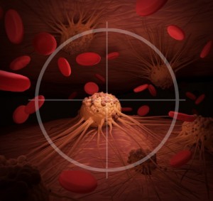
Cancer is the second most common cause of death in developed countries. Statistical data from the last few years indicate that cancer is responsible for about a quarter of all deaths. Although the number of cases continues to grow, diagnostic methods are improving, and if the cancer is detected early, it can often be effectively treated. This is partly because scientists and doctors from around the world are becoming more knowledgeable in the field of oncology and they discover more effective ways to diagnose and treat cancer. Researchers from Warsaw University of Life Sciences had significant contribution to the development of knowledge about cancer. At WULS-SGGW, several major projects aimed at combating cancer are carried out.
New hope
Currently, the most promising direction of research into effective cancer therapies is immunooncology. It’s a new strategy of using the patient’s immune system. Time will tell whether the results of research conducted by scientists around the world will prove to be a breakthrough in medicine, or whether they will be used to develop methods for treating just some types of cancer. At the Faculty of Veterinary Medicine at WULS-SGGW, there are two research teams that work on the projects from the field of immunooncology.
Backpacks with bomb against cancer
Magdalena Król, Dr. hab., professor at WULS-SGGW, has in 2016 received a prestigious grant of the European Research Council (ERC). Its value is over 1.4 million Euro. It will be spent on a five-year research project during which the researcher and her team will try to explain a biological phenomenon that she had discovered. She noted namely that certain immune system cells transfer a specific group of proteins to tumor cells and she came up with the idea of using this knowledge in oncology practice. Professor Król intends to use this mechanism to precisely deliver cancer-destroying drugs directly to the tumor. It’s as if the cells of the immune system of the patient were wearing backpacks with explosives which aim at the cancer – says Magdalena Król.
Currently, only 2% of the drugs applied to patients get into the solid tumor – and they only get to the sites located close to blood vessels. The rest of the drugs cause devastation in the body while the undamaged cancer cells, hidden in hypoxic and non-vascularised places, multiply causing metastasis and recurrence. The use of physiological processes of the body: the cells of the immune system that could deliver drugs to places inaccessible to conventional therapy could significantly increase the chances of curing the sick and solve one of the biggest problems of oncology – metastasis. If the research hypothesis is confirmed, it might be a breakthrough in the treatment of cancer.
The possibility that the discovery of prof. Magdalena Król will become a milestone in the treatment of cancer is high. As many as 20% of projects implemented in the framework of the ERC grants has led to a real breakthrough in science, and the results of 50% of the projects contributed to significant progress in the area. It is not easy to obtain financing from the European Research Council. Detailed applications are evaluated by a body of eminent scientists and experts from the specific field.
Trained lymphocytes
Also Kinga Majchrzak, Ph.D., works in the area of immunooncology at the Faculty of Veterinary Medicine. Her first research on cancer was related to breast cancer in dogs. After defending her dissertation, she began working with Dr. Chrystal Paulos, staying at her lab at the Medical University of South Carolina in the United States a total of two years. Dr. Paulos studies the use of T cells in adoptive cell therapy including melanoma and pancreatic cancer. During her academic stay, Dr. Majchrzak investigated how Th17 cells affect the treatment of melanoma in mice. The aim of her laboratory work was to modify their properties so that they are most effective in combating cancer.
I was working on blocking signaling pathways in lymphocytes Th17 given to sick mice and checking in which case the performance of lymphocytes is higher. First, I blocked in vitro signaling pathways in cells, and then I gave these modified cells to mice. Test result was very interesting – in laboratory breeding, the lymphocytes with blocked signaling pathways were weaker than the ones whose pathways were not modifed. However, when they were given in vivo to the organism of mice, they proved to be more effective than the control cells. Mice that received Th17 cells treated with the two inhibitors were in the best condition; some of them recovered, and in the remaining mice, the tumor growth was inhibited. The research conducted by the scientists in the United States is an important step towards improving the effectiveness of treatment with T-cell. The point is that therapy should have as few side effects as possible, such as cytokine storm, or autoimmune reaction – says Kinga Majchrzak.
After returning to Poland, she won a grant to conduct her own experiments in the field of immunoecology. She is just starting a study on Th17 lymphocytes and their interactions with other cells in dogs. Her research plans, however, have a much broader scale. Dr. Majchrzak wants to cooperate with veterinarians and oncologists to conduct clinical trials using modified cells in dogs with cancer. In my opinion this is a very good solution. We can carry out clinical trials of new treatments and observe how a living organism reacts. Such studies would have much higher cognitive value than the ones performed on mice. Dogs suffer from similar cancers as people and the disease is almost the same. Als, dogs would benefit this way by getting a chance to recover. The results of these studies would benefit both veterinary and medicine – she explains.
At the Warsaw University of Life Sciences, the next team engaged in research on cancer is being formed. Dr. Kinga Majchrzak was recently joined by a PhD student, MSc Joanna Bujak, who came up with the idea that the same cells could be used to investigate calcium and sodium ion channels for sodium and calcium. I will work on, among other,, TRPM5 protein, which is known to be blocked by low pH. Lower pH distinguishes the tumor from a healthy tissue. I want to see if this mechanism is responsible for the fact that the tumor protects itself from the action of lymphocytes, even if they fail to locate cancer cells – explains Joanna Bujak.
In the interest of animals
Animals play a very important role in the cancer research because new therapies are tested on them. However, the majority of research – even if at a certain stage performed in veterinary laboratories – is carried out in order to develop effective treatments for humans. It is true that the latest medical knowledge can be also used in veterinary medicine, but it doesn’t benefit the veterinary patients in all cases. The cost of oncological diagnostics of animals often exceeds the financial capacity of their owners. However, there are scientists who want to change this situation. One of them is Dr. hab. Rafal Sapierzyński, professor at the Warsaw University of Life Sciences, Department of Animal Pathology, Faculty of Veterinary Medicine, who works on the epidemiology and detection of tumors in animals, mainly dogs and cats. The Professor develops inexpensive and minimally invasive tests that will enable the the largest set of information about the condition of the animal. This knowledge will help doctors to quickly diagnose the patients, to make prognosis and to specify the method and cost of treatment. As a result, more pet owners will be able to decide to treat their pets.
Graphene and platinum Trojan horse
Researchers from the Department of Nutrition and Animal Biotechnology of WULS-SGGW are getting good results in the fight against cancer. For the last ten years, research using a variety of nanostructures in the area of broadly defined biology, especially in medicine, has been carried out under the guidance of prof. Ewa Sawosz- Chwalibog. The ultimate goal of the research is to explore and construct innovative nano-molecules that could have anti-cancer effects.
Most of the research is focused on the fight against one of the most dangerous cancers, which is glioblastoma grade IV. In the Department of Nanobiotechnology, the team led by Dr. Marta
Grodzik[1] conducted the world’s first study on the use nano-platinum. Although platinum-based drugs in medicine are popular and quite effective, they also cause numerous serious side effects.
Research shows that the toxicity of nano-platinum is much lower and its use in the treatment is safer for the patient.
Similar research carried out at the same time on graphene showed that the graphene oxide flakes are effective in the treatment of brain tumors and breast cancer. Graphene is a form of carbon with intricate designs. Joining graphene with oxygen groups increases its biocompatibility with the body, but does not change the lethal action on cancer cells.
Researchers from the WULS-SGGW went a step further and combined graphene oxide flakes with platinum nanoparticles. The resulting graphene-platinum may be used as a therapeutic measure injected directly into the tumor and surrounding tissue. As coal is recognized as friendly molecule, the obtained material is freely located in the tumor cells and just as the Trojan horse, gradually releases ions of platinum, which damage the DNA of cancer cells. As the nano- molecule only works at the injection site, there is low risk that the active substance enters into reactions with the healthy cells of the body.
Although the pilot study provides hope for a long-awaited breakthrough in the fight against glioblastoma, still lot more testing is needed to translate this knowledge into clinical practice.
From the point of view of Artificial Intelligence
Determining the stage of cancer is sometimes difficult even for experienced pathologists. Perhaps artificial intelligence will help them soon in their practice. At the Faculty of Applied Informatics and Mathematics of WULS-SGGW a tem of researchers led by Dr. hab. Eng. Michał Kruk works on programming automated computer diagnostics. Properly prepared neural networks are trained to recognize histopathomorphology images, that is microscopic images showing the specimen collected during the biopsy. Even the most developed computer programs can not replace a physician pathologist. However, in extreme cases or unclear situations, they are often more accurate than doctors. So they can be a very helpful tool in the hands of an experienced specialist – says Michal Kruk.
Researchers at the Warsaw University of Life Sciences have worked with Military Medical Institute in Warsaw for years. They have already had some successes – they were able to, among other, teach artificial intelligence to recognize clear-cell renal cancer. The application that was created is used to aid diagnosis in the Department of Pathology in the Military Medical Institute. Currently, researchers conduct studies on milk ducts cancer in human breast. The program that is created will recognize tumors on the basis of images and it will inform whether they are cystic (non-invasive) or already invasive. The implementation of this project will take 2-3 years.
Anna Ziółkowska
Cooperation: Katarzyna Wolanin
[1]The team composed of: Ewa Sawosz-Chwalibóg, Marta Grodzik, Mateusz Wierzbicki, Marta Kutwin, Sławomir Jaworski, Anna Hotowy, Barbara Strojny, Natalia Kurantowicz


